Wednesday, September 23, 2020

Excellent intervention points against SARS-CoV-2 infections discovered.

" Our findings provide a direct structural link between LA, COVID-19 pathology and the virus itself and suggest that both the LA-binding pocket within the S protein and the multi-nodal LA signaling axis, represent excellent therapeutic intervention points against SARS-CoV-2 infections."


Abstract

COVID-19, caused by severe acute respiratory syndrome-coronavirus-2 (SARS-CoV-2), represents a global crisis. Key to SARS-CoV-2 therapeutic development is unraveling the mechanisms driving high infectivity, broad tissue tropism and severe pathology. Our 2.85 Å cryo-EM structure of SARS-CoV-2 spike (S) glycoprotein reveals that the receptor binding domains (RBDs) tightly bind the essential free fatty acid (FFA) linoleic acid (LA) in three composite binding pockets. The pocket also appears to be present in the highly pathogenic coronaviruses SARS-CoV and MERS-CoV. LA binding stabilizes a locked S conformation giving rise to reduced ACE2 interaction in vitro. In human cells, LA supplementation synergizes with the COVID-19 drug remdesivir, suppressing SARS-CoV-2 replication. Our structure directly links LA and S, setting the stage for intervention strategies targeting LA binding by SARS-CoV-2.

SIGN UP FOR THE SCIENCE eTOC

Get the latest issue of Science delivered right to you!

Seven coronaviruses are known to infect humans. The four endemic human coronaviruses OC43, 229E, HKU1 and NL63 cause mild upper respiratory tract infections while pandemic virus SARS-CoV-2, and earlier SARS-CoV and MERS-CoV, can cause severe pneumonia with acute respiratory distress syndrome, multi-organ failure, and death (12).

SARS-CoV-2 has acquired functions that promote its harsh disease phenotype. SARS-CoV-2 causes severe inflammation and damage to endothelial cells in the heart, kidneys, liver and intestines, suggestive of a vascular infection rather than a purely respiratory disease (34). The attachment of SARS-CoV-2 to a host cell is initiated by the spike protein trimer (S), which decorates the outer surface of the virus, binding to its cognate receptor angiotensin-converting enzyme 2 (ACE2), with higher affinity than SARS-CoV (57). A S1/S2 polybasic furin protease cleavage site distinguishes SARS-CoV-2 from SARS-CoV or other closely related bat coronaviruses and serves to stimulate entry into host cells and cell-cell fusion (589). Inside the host cell, human coronaviruses remodel the lipid metabolism to facilitate virus replication (10). Infection by SARS-CoV-2 triggers an unusually impaired and dysregulated immune response (11) and a heightened inflammatory response (12) working in synergy with interferon production in the vicinity of infected cells to drive a feed-forward loop to up-regulate ACE2 and further escalate infection (13).

In the search for additional functions that contribute to the pathology of infection, we determined the structure of the SARS-CoV-2 S glycoprotein by cryo-EM (Fig. 1). We produced SARS-CoV-2 S as a secreted trimer (14) in MultiBac (15) baculovirus-infected Hi5 insect cells (fig. S1) (16). Highly purified protein was used for cryo-EM data collection (fig. S2 and table S1). After 3D classification and refinement without applying symmetry (C1) we obtained a 3.0 Å closed conformation from 136,405 particles and a 3.5 Å open conformation with one receptor-binding domain (RBD) in the up position from 57,990 particles (figs. S2 and S3). C3 symmetry was applied to the closed conformation particle pool yielding a 2.85 Å map (Fig. 1A and figs. S2 and S3).

Fig. 1 Cryo-EM structure of SARS-CoV-2 spike linoleic acid complex.

(A) Cryo-EM density of spike trimer (left). Monomers in cyan, green and pink, respectively. The structure in a cartoon representation in a front (middle) and top view (right). Bound LA illustrated as spheres colored in orange. One LA-binding pocket is boxed in red. (B) Composite LA-binding pocket formed by adjacent RBDs. Tube-shaped EM density is shown. (C) LC-MS of purified S. Chemical structure and molecular weight of LA (top), C4 column elution profile (middle) and ESI-TOF of wash solution (gray) and C4 peak elution fraction (black) with peak molecular weight indicated (bottom). (D) Hydrophobic LA-binding pocket in a surface representation illustrating excellent fit of bound LA (orange, sticks and balls representation). Blue and red indicate positive and negative surface charge, respectively. (E) LA interactions with amino acids in the binding pocket. The acidic LA headgroup is in the vicinity of an arginine (408) and a glutamine (409).

The structure of S displays the characteristic overall shape observed for coronavirus S proteins in the closed and open conformations (1719) with the closed form (~70%) predominating in our data set (Fig. 1A and figs. S2 to S4). Model building of the closed form revealed additional density in the RBDs in our structure (Fig. 1B). The tube-like shape of this density was consistent with a fatty acid, with size and shape similar to LA bound to other proteins (Fig. 1B and fig. S5) (2021). Liquid chromatography coupled ESI-TOF mass spectrometry (LS-MS) analysis confirmed the presence of a compound with the molecular weight of LA in our purified sample (Fig. 1C).

Hallmarks of FFA-binding pockets in proteins are an extended “greasy” tube lined by hydrophobic amino acids which accommodates the hydrocarbon tail, and a hydrophilic, often positively charged anchor for the acidic headgroup of the FFA. In our structure, a hydrophobic pocket mostly shaped by phenylalanines forms a bent tube into which the LA fits well (Fig. 1, D and E). The anchor for the headgroup carboxyl is provided by an arginine (R408) and a glutamine (Q409) from the adjacent RBD in the trimer, giving rise to a composite LA-binding site (Fig. 1E). We confirmed the presence of LA in all three binding pockets in the S trimer in the unsymmetrized (C1) closed structure (fig. S6). Similarly, masked 3D classification focusing on the RBD domains could not identify any unoccupied pockets (fig. S7).

Our S construct contains alterations as compared to native SARS-CoV-2 S namely addition of a trimerization domain and deletion of the polybasic cleavage site, neither of which alter S conformation appreciably (1417) (fig. S8). Glycosylation sites are located away from the LA-binding pocket and largely native in our structure (717) (table S2). Thus, neither mutations nor glycosylation are likely to impact the LA-binding pocket. We compared S and RBD produced in insect cells with mammalian produced S to identify any potential influence of differences in glycosylation on ACE2 binding by competition enzyme-linked immunosorbent assay (ELISA) (Fig. 2A). All three reagents bound ACE2 efficiently. We further confirmed ACE2 binding by S using SEC with purified proteins (Fig. 2B). The LA-binding pocket and the receptor binding motif (RBM) are distal and non-overlapping (Fig. 2C). Notably, in the LA-bound S the RBM is ordered and buried at the interface between RBDs while it was disordered in previously described SARS-CoV-2 S cryo-EM structures (717).

Fig. 2 Functional characterization of LA-bound SARS-CoV-2 spike.

(A) Insect cell (Hi5) expressed spike (dark blue bars), insect cell expressed RBD (light blue bars) and mammalian (HEK293) expressed spike (white bars) in competition ELISAs utilizing immobilized ACE2. Error bars: standard deviations, three replicates. (B) LA-bound SARS-CoV-2 spike protein interaction with ACE2 was analyzed by size exclusion chromatography evidencing complex formation. SEC profiles (left) for ACE2 (yellow, III), LA-bound spike (green, II) and a mixture of ACE2 and LA-bound spike (orange, I) are shown. Peak fractions (I.-III.) were analyzed by SDS-PAGE evidencing the expected proteins (right). (C) LA-bound S glycoprotein trimer top view with RBDs shown in cyan, green and pink respectively (left). In each RBD subunit the motif responsible for ACE2 binding (RBM) is in red, and LA is shown as spheres colored in orange. A close-up view into the cyan RBD (right) shows that the RBM is fully ordered, and LA and RBM are not in direct contact. (D) SPR analysis of the binding of LA-bound spike trimer (orange curves) and apo spike trimer (green curves) to immobilized ACE2. Apo spike and LA-bound spike were diluted to concentrations of 40 nM and 160 nM, respectively. Black lines correspond to a global fit using a 1:1 binding model. (E) Synergistic effect of LA and remdesivir on SARS-CoV-2 viral replication. Effects of varying doses of remdesivir ± 50 μM LA on virus infection are shown. Human Caco-2 ACE2+ cells were infected with SARS-CoV-2 and then treated with varying doses of remdesivir ± 50 μM LA. At 96 hours post-infection, cells were fixed and infected cells detected by immunofluorescence assay using an anti-N antibody (green). Cell nuclei were stained by DAPI (blue). Representative images corresponding to the remdesivir dose range 20 – 200 nM are shown. (F) The amount of extracellular virus present in wells (n = 3) at the dose combinations shown was determined by qRT-PCR.

SARS-CoV-2 S can also adopt an open conformation (fig. S4) which is compatible with binding ACE2. In previous apo S cryo-EM structures about 60-75% of the S trimers were in the open conformation (717), contrasting our observation of ~70% in the closed conformation. This could be due to LA stabilizing the closed conformation, and if so LA would be expected to reduce ACE2 binding. We performed surface plasmon resonance (SPR) experiments with biotinylated ACE2 immobilized on a streptavidin-coated chip (Fig. 2D and fig. S9). We first determined the KD of the RBD/ACE2 interaction to validate our assay. Our value (26 nM, fig. S9C) is in good agreement with previous studies (44 nM (22)) obtained by SPR with the RBD immobilized and ACE2 as analyte. Apo S was prepared by applying Lipidex, the established method for removing lipids from lipid-binding proteins (23). A KD of 0.7 nM was obtained for the apo S/ACE2 interaction (fig. S9A). For the LA-bound S/ACE2 interaction we obtained a KD of 1.4 nM (fig. S9B). We consistently obtained a markedly reduced resonance unit (RU) signal for LA-bound S as compared to apo S at the same concentrations (Fig. 2D and fig. S9, A and B). This correlates with the apo state having a higher percentage of S trimers in the open, ACE2-accessible conformation.

We characterized the affinity of the LA interaction both experimentally and computationally. Our SPR assays utilizing immobilized RBD yielded a binding constant of ~41 nM exhibiting a slow off-rate, consistent with tight binding of LA (fig. S10). Repeated molecular dynamics simulations of the entire locked LA-bound spike trimer (3 × 100 ns) using GROMACS-2019 (24) corroborated the persistence of stable interactions between LA and the spike trimer (movies S1 and S2). The affinity of LA binding to the spike trimer will likely be higher than to the RBD alone, taking into account polar headgroup interactions with R408 and Q409 of the adjacent RBD (Fig. 1E). The resolution of the RBDs in our open S cryo-EM structure was insufficient to either assign, or rule out, a ligand-bound pocket (fig. S3). However, the slow off-rate observed with the RBD monomer (fig. S10) suggests that LA binding could be maintained when the S trimer transiently converts into the open conformation. This is supported by our observation that LA was retained during S purification in spite of S trimers adopting the open form ~30% of the time (fig. S2) and by our MD simulations with a modeled ligand-bound open spike trimer (movie S3) in which all three LAs remained bound over 500 ns.

Next, we investigated the effect of LA in experiments using live SARS-CoV-2 virus to infect human epithelial cells. Remdesivir is an RNA-dependent RNA polymerase inhibitor and the first anti-viral drug showing benefit in the treatment of COVID-19 in clinical trials, albeit with considerable side effects at the doses required (25). LA supplementation at 50-100 μM concentrations was previously shown to affect coronavirus entry and replication (10). We administered remdesivir at 20, 64 and 200 nM concentration, supplementing with 50 μM LA (Fig. 2E). Our results revealed synergy, with the dose of remdesivir required to suppress SARS-CoV-2 replication markedly reduced by adding LA (Fig. 2, E and F).

We superimposed our LA-bound structure on previous SARS-CoV-2 apo S structures in the closed conformation (717) and identified a gating helix located directly at the entrance of the binding pocket (Fig. 3, A to C). This gating helix, comprising Ty365 and Tyr369, is displaced by about 6 Å when LA is bound, thus opening the pocket (Fig. 3, A and B). In the apo SARS-CoV-2 S trimer (717), a gap between adjacent RBDs places the hydrophilic anchor residues ~10 Å away from the position of the LA headgroup (Fig. 3C). Upon LA binding, the adjacent RBD in the trimer moves toward its neighbor, and the anchor residues Arg408 and Gln409 lock down on the headgroup of LA (Fig. 3, C and D). Overall, this results in a compaction of trimer architecture in the region formed by the three RBDs giving rise to a locked S structure (Fig. 3D and movie S4).

Fig. 3 Comparison of LA-bound and apo S structures.

(A) Superimposition of LA-bound SARS-CoV-2 (cyan RBD) and the ligand-free “apo” RBD (gray) (PDBID 6VXX (7)). The gating helix at the entrance of the hydrophobic pocket moves by 6 Å in the presence of LA. Tyr365 and Tyr369 swing away avoiding clashes with LA (orange). Black arrows indicate the rearrangements. (B) The same structure as panel A rotated by 90° as indicated, showing the entrance of the hydrophobic pocket. (C) Formation of a composite LA-binding pocket by two adjacent RBDs in LA-bound S involves a ~5 Å movement of RBD2 (green) toward RBD1 (cyan) compared to apo S (gray). (D) Superimposition of the RBD trimer of apo S (gray) and LA-bound S (RBD1 cyan, RBD2 green, RBD3 pink, LA orange) is shown (left). The individual RBD trimers are depicted for LA-bound S (right, top) and apo S (right, bottom) with RBDs boxed in black, highlighting the compaction of RBDs in the LA-bound S structure.

We investigated whether the LA-binding pocket is conserved in the seven coronaviruses that infect humans (Fig. 4A and table S3). Sequence alignment shows that all residues lining the hydrophobic pocket and the anchor residues (Arg408/Gln409) in SARS-CoV-2 are fully conserved in SARS-CoV (Fig. 4A). Structural alignment of LA-bound RBDs within the trimer of SARS-CoV-2 and “apo” SARS-CoV RBDs (19) reveals that the LA-binding pocket is present in SARS-CoV. The greasy tube is flanked by a gating helix as in SARS-CoV-2, with Arg395/Gln396 of SARS-CoV positioned 10 Å and 11 Å from the entrance, respectively, virtually identical to apo SARS-CoV-2 (Figs. 3C and 4B). In MERS-CoV, the gating helix and hydrophobic residues lining the pocket are also present. Tyr365, Tyr369 and Phe374 are substituted by likewise hydrophobic leucines and a valine, respectively (Fig. 4, A and C) (19). The Arg408/Gln409 pair is not conserved, however, we identify Asn501/Lys502 and Gln466 as potential anchor residues, located on a β-sheet and an α-helix within the adjacent RBD, up to 11Å away from the entrance (Fig. 4C). Thus, the greasy tube and hydrophilic anchor appear to be present in MERS-CoV, suggesting convergent evolution. In HCoV OC43, gating helix and hydrophobic residues lining the pocket are largely conserved, while Tyr365, Tyr369 and Phe374 are replaced by methionines and alanine, respectively (Fig. 4A) (18). Arg413 is located on the same helix as Arg408/Gln409 in SARS-CoV-2 and could serve as a hydrophilic anchor (Fig. 4D). No gap exists in this presumed “apo” form structure between the RBDs which appear already in the locked conformation (Fig. 4D and fig. S11) (18). In HCoV HKU1, the hydrophobic residues are again largely conserved, but a charged residue (Glu375) is positioned directly in front of the entrance, obstructing access for a putative ligand (Fig. 4E) (26). The RBDs of HCoVs 229E and NL63 adopt a very different fold (fig. S13) (2728), and many of the LA-binding residues are not present (Fig. 4A), hampering predictions of a binding site for fatty acids.

Fig. 4 Human coronavirus RBD architectures.

(A) Alignments of the seven CoV strains that can infect humans, highlighting conserved residues. Residues lining the hydrophobic pocket are underlaid (cyan). Gating helix residues are marked (purple). Residues positioned to interact with the LA polar headgroup are underlaid in green. Glu375 in HKU1 is underlaid in red (cf. panel E). (B) Superimposition of RBD1 of LA-bound SARS-CoV-2 (RBD2 in green) with RBD1 of ligand free “apo” SARS-CoV (RBD1 and RBD2 magenta, PDBID 5X58 (19)) indicates a conservation of the composite binding pocket. (C) Superimposition of RBD1 of LA-bound SARS-CoV-2 (RBD2 is omitted for clarity) with RBD1 of MERS-CoV (RBD1 and RBD2 forest green, PDBID 5X5F (19)). (D) Superimposition of RBD1 of LA-bound SARS-CoV-2 (RBD2 in green) with RBD1 of OC43 (RBD1 and RBD2 purple, PDBID 6NZK (18)). (E) Superimposition of LA-bound SARS-CoV-2 RBD with HKU1 RBD (brown, PDBID 5GNB (26)). LA is omitted in SARS-CoV-2 RBD for clarity.

In summary, we find four molecular features mediating LA binding to SARS-CoV-2, and potentially also SARS-CoV and MERS-CoV S proteins: a conserved hydrophobic pocket, a gating helix, amino acid residues pre-positioned to interact with the LA carboxy headgroup, and loosely packed RBDs in the “apo” form. In contrast, in each of the four common circulating HCoVs, it appears that one or more of these four architectural prerequisites are lacking in the S protein structures (Fig. 4 and figs. S11 and S12). LA binding to SARS-CoV-2 S triggers a locking down of the hydrophilic anchor and a compaction of the RBD trimer (Fig. 3, C and D). In addition to stabilizing the closed conformation, this could also help stabilize the S1 region comprising the N-terminal domain and the RBD. The RBM, central to ACE2 binding, appears to be conformationally preorganized in our structure (Fig. 2C) indicating a generally more rigid RBD trimer when LA is bound. While direct crosstalk in between the LA-binding pocket and the RBM is not apparent from our structure (Fig. 2C), the conformational changes in the RBD trimer (Fig. 3) could impact ACE2 docking and infectivity as indicated by our SPR assays showing reduced levels of S binding in the presence of LA (Fig. 2D). The S protein’s tight binding of LA originates from a well-defined size and shape complementarity afforded by the pocket (Fig. 1, B and D). The LA-binding pocket thus presents a promising target for future development of small molecule inhibitors that, for example, could irreversibly lock S in the closed conformation and interfere with receptor interactions. It is noteworthy in this context that a fatty acid binding pocket was exploited previously to develop potent small molecule anti-viral drugs against rhinovirus, locking viral surface proteins in a conformation incompatible with receptor binding (2930). These anti-virals were successful in human clinical trials (3132).

A recent proteomic and metabolomic study of COVID-19 patient sera showed continuous decrease of FFAs including LA (33). Lipid metabolome remodeling is a common element of viral infection (3435). For coronaviruses, the LA to arachidonic acid metabolic pathway was identified as central to lipid remodeling (10). We hypothesize that LA sequestration by SARS-CoV-2 could confer a tissue-independent mechanism by which pathogenic coronavirus infection may drive immune dysregulation and inflammation (3537). Our findings provide a direct structural link between LA, COVID-19 pathology and the virus itself and suggest that both the LA-binding pocket within the S protein and the multi-nodal LA signaling axis, represent excellent therapeutic intervention points against SARS-CoV-2 infections.

Supplementary Materials

science.sciencemag.org/cgi/content/full/science.abd3255/DC1

Materials and Methods

Figs. S1 to S12

Tables S1 to S3

References (4163)

MDAR Reproducibility Checklist

Movies S1 to S4

https://creativecommons.org/licenses/by/4.0/

This is an open-access article distributed under the terms of the Creative Commons Attribution license, which permits unrestricted use, distribution, and reproduction in any medium, provided the original work is properly cited.

References and Notes

  1. To produce spike glycoprotein, we used here baculovirus-infected insect cells (Hi5) cultured in ESF921 media which contains cod liver oil as a nutrient supplement (Corey Jacklin, Expression Systems, personal communication). Cod liver oil comprises hundreds of FFAs including LA (38) which could be a possible source of the fatty acid. In parallel to our work, cryo-EM structures were determined of spike expressed in a mammalian system (HEK293) in serum and protein-free media (3940). The tube-shaped density we identified in our study is also present in those structures (but was not assigned or interpreted). We conclude that LA binding in the SARS-CoV-2 S pocket is thus not dependent on the expression system or media used.
Acknowledgments: We thank all members of the Berger and Schaffitzel teams as well as Gunjita Singh, Yohei Yamauchi and David Matthews (University of Bristol, UK) for their assistance. We thank Florian Krammer (Icahn School of Medicine, USA) for kindly sharing expression plasmids. We thank Veronica Chang and Radu Aricescu (MRC-LMB, UK) for kind gift of S expressed in HEK293. We are indebted to Adam Finn (Bristol UNCOVER Group and Children’s Vaccine Centre, Bristol Medical School), Jeremy Tavaré (School of Biochemistry, Bristol), Kathleen Gillespie (Diabetes and Metabolism Unit, Southmead Hospital, Univ. of Bristol) and Donald Fitzgerald MD (Quest Imaging Medical Associates, USA) for helpful discussions and careful reading of the manuscript. We thank Simon Burbidge, Thomas Batstone and Matt Williams for computation infrastructure support. We would like to thank the Advanced Computing Research Centre (ACRC) at the University of Bristol for access to BlueCryo, BlueCrystal Phase 4 and BlueGEM, and the UK HECBioSim for access to the UK supercomputer, ARCHER. We are particularly grateful to Thiru Thangarajah (Genscript) for early access to Genscript’s cPass SARS-CoV-2 Neutralization Antibody Detection/Surrogate Virus Neutralization Test Kit (L00847). We thank Sebastian Fabritz and the Core Facility for Mass Spectrometry at the Max Planck Institute for Medical Research for their support on MS measurements. Funding: This research received support from the Elizabeth Blackwell Institute for Health Research and the EPSRC Impact Acceleration Account EP/R511663/1, University of Bristol, from BrisSynBio a BBSRC/EPSRC Research Centre for synthetic biology at the University of Bristol (BB/L01386X/1) (to I.B., A.J.M., D.K.S.) and from the BBSRC (BB/P000940/1) (to C.S. and I.B.). This work received generous support from the Oracle Higher Education and Research program to enable cryo-EM data processing using Oracle’s high-performance public cloud infrastructure (https://cloud.oracle.com/en_US/cloud-infrastructure) and the EPSRC through a COVID-19 project award via HECBioSim to access ARCHER (A.J.M.). We acknowledge support and assistance by the Wolfson Bioimaging Facility and the GW4 Facility for High-Resolution Electron Cryo-Microscopy funded by the Wellcome Trust (202904/Z/16/Z and 206181/Z/17/Z) and BBSRC (BB/R000484/1). The authors would like to acknowledge support of the University of Bristol's Alumni and Friends, which funded the ImageXpress Pico Imaging System. O.S. acknowledges support from the Elisabeth Muerer Foundation, the Max Planck School Matter to Life and the Heidelberg Biosciences International Graduate School. A.D.D. is supported by the United States Food and Drug Administration (HHSF223201510104C) and UK Research and Innovation/Medical Research Council (MRC) (MR/V027506/1). M.K.W is supported by MRC grants MR/R020566/1 and MR/V027506/1 (awarded to A.D.D). A.J.M. is supported the British Society for Antimicrobial Chemotherapy (BSAC-COVID-30) and the EPSRC (EP/M022609/1, CCP-BioSim). I.B. acknowledges support from the EPSRC Future Vaccine Manufacturing and Research Hub (EP/R013764/1). C.S. and I.B. are Investigators of the Wellcome Trust (210701/Z/18/Z; 106115/Z/14/Z). Author contributions: C.S. and I.B. conceived and guided the study. F.G., K.G. and J.C. produced, purified and analyzed sample, K.G. carried out biochemical experiments, S.K.N.Y. and U.B. prepared grids and collected EM data, S.K.N.Y., U.B., K.G and C.T. carried out image analysis and model building. A.D.D., M.K.W. and R.M. performed all live virus CL3 work and analyzed data. D.K.S. and A.J.M. performed all MD simulations. O.S. and J.S. performed and interpreted mass spectrometry. C.T., K.G., D.F., I.B. and C.S. interpreted results. D.F., I.B. and C.S. wrote the manuscript with input from all authors. Competing interests: The authors declare competing interests. I.B and F.G. report shareholding in Imophoron Ltd. unrelated to this Correspondence. I.B. and D.F. report shareholding in Geneva Biotech Sàrl related to this Correspondence. Patent applications describing methods and material compositions based on the present observations have been filed. Data and materials availability: Datasets generated during the current study have been deposited in the Electron Microscopy Data Bank (EMDB) under accession numbers EMD-11145 (C3 closed conformation), EMD-11144 (C1 closed conformation) and EMD-11146 (open conformation), and in the Protein Data Bank (PDB) under accession numbers: 6ZB5 (C3 closed conformation) and 6ZB4 (C1 closed conformation). Reagents are available from I.B. and C.S. under a material transfer agreement with the University of Bristol. This work is licensed under a Creative Commons Attribution 4.0 International (CC BY 4.0) license, which permits unrestricted use, distribution, and reproduction in any medium, provided the original work is properly cited. To view a copy of this license, visit https://creativecommons.org/licenses/by/4.0/. This license does not apply to figures/photos/artwork or other content included in the article that is credited to a third party; obtain authorization from the rights holder before using such material.

View Abstract

Recommended articles from TrendMD

  1. Structural basis for the recognition of the SARS-CoV-2 by full-length human ACE2
    Renhong Yan et al., Science,  2020
  2. A highly conserved cryptic epitope in the receptor-binding domains of SARS-CoV-2 and SARS-CoV
    Meng Yuan et al., Science,  2020
  3. Structural basis for neutralization of SARS-CoV-2 and SARS-CoV by a potent therapeutic antibody
    Zhe Lv; Yong-Qiang Deng; Qing Ye; Lei Cao; Chun-Yun Sun; Changfa Fan; Weijin Huang; Shihui Sun; Yao Sun; Ling Zhu; Qi Chen; Nan Wang; Jianhui Nie; Zhen Cui; Dandan Zhu; Neil Shaw; Xiao-Feng Li; Qianqian Li; Liangzhi Xie; Youchun Wang; Zihe Rao; Cheng-Feng Qin; Xiangxi Wang, Science 
  4. A neutralizing human antibody binds to the N-terminal domain of the Spike protein of SARS-CoV-2
    Xiangyang Chi et al., Science 
  5. Cryo-EM structure of the 2019-nCoV spike in the prefusion conformation
    Daniel Wrapp et al., Science,  2020

Tuesday, September 15, 2020

WHY CANADA, WHY?

 WHY CANADA...WHY?

ATTENTION:  Foreign Affairs Minister François-Philippe Champagne it's time for Canada to stop dealing in forest destruction.

 THE AMAZON IS BURNING AND SADLY CANADA IS PARTLY RESPONSIBLE! THE QUESTION IS WHY CANADA...WHY?

 ONE POINT EIGHT BILLION DOLLARS IS THE AMOUNT CANADA WILL PAY FOR BRAZILIAN BEEF. IT IS THE REASON BRAZILIAN FARMERS AND RANCHERS ARE BURNING THE AMAZON FOREST. THEY IGNORANTLY BELIEVE THEY ARE MAKING ROOM FOR CATTLE RANCHING AND FUTURE PROFITS  BUT IN REALITY THEY ARE DESTROYING MILLIONS OF PLANT AND ANIMAL SPECIES AND MURDERING INDIGENOUS PEOPLE! AND THE ONLY CONSEQUENCE  BURNING THE AMAZON FOREST WILL CREATE? DESERTIFICATION! 

 THE AMAZON FOREST CREATES PRECIPITATION AND WITHOUT THE FOREST THE AREA WILL DRY UP. THIS SCIENCE BASED FACT HAS NOT YET ENTERED THE SMALL MINDS PRESENTLY BURNING THE FOREST AND NEITHER HAS IT ENTERED INTO THE GREED FILLED CANADIAN NO-BRAINS PRESENTLY CREATING A FREE TRADE DEAL WITH BRAZIL. A FREE TRADE DEAL THAT IS MINUS ONE IMPORTANT CONDITION...PROTECT THE AMAZON RAIN FOREST! AND WHAT DO CANADIAN CATTLE RANCHERS AND FARMERS HAVE TO SAY ABOUT THIS DEAL?               

Hi Nelson,

I’m not going to lie - last week was tough. And if your newsfeeds are anything like mine, it was tough for you too. 

Images of sickeningly orange skylines and smoke-filled air poured in from across the globe - Brazil, Greece, Bulgaria, Argentina, California.

When it comes to the fires in the Amazon right now, however, I have hope because Canada has a direct role to play in putting them out. But we need to act fast.

Right now, our government is planning to proceed with negotiations for a Canada-Mercosur free trade deal with Brazil. And Brazil’s big agribusiness is celebrating the move. Why? Because the deal is expected to increase Brazil’s meat exports to Canada by as much as $1.8 BILLION annually. [1]

And yup - you guessed it. The production of cheap meat is the lead driver of the Amazon fires!

Will you join us in sending a message to Canada’s Minister of Foreign Affairs Francois-Philippe Champagne to immediately halt the Canada-Mercosur free trade negotiations and protect the Amazon?

Fires in the Amazon rainforest are not natural. They are deliberately set by land-grabbers and ranchers to expand the land used for cattle grazing and industrial agriculture production.[2] Brazil’s President Jair Bolsonaro has fueled the problem by dismantling environmental laws, slashing funding for environmental protection agencies and demolishing Indigenous land rights.

This has given free reign to big agro to clear and burn the forest. There has even been a surge in murders of Indigenous forest guardians as land-grabbers move in on Indigenous lands. [3]

Canada cannot be complicit in the destruction of the Earth’s vital forests and the violation of Indigenous rights. But our Foreign Affairs Minister needs to hear it from all of you before the trade deal goes through.

There is so much at stake - every person on earth depends on the Amazon to help regulate our climate. The rainforest sucks carbon from the air and stores it in billions of trees. It produces a “flying river” of water vapour that distributes rain across South America and impacts weather in other continents. [4]

If deforestation continues, the surviving forest would no longer be able to produce its own rainfall and quickly transform into an arid savannah – killing off species and releasing massive amounts of carbon into the atmosphere.

But this won’t move Brazil to act. Thus far, the only thing that has influenced Bolsonaro is intense international pressure from governments threatening to cut business ties unless the Amazon is protected. [5]

France and Germany have already halted further movement to ratify Europe's free trade deal with the Mercosur bloc. [6] What are we waiting for?  

It's time for Canada to halt this deal too. Tell Foreign Affairs Minister François-Philippe Champagne it's time for Canada to stop dealing in forest destruction.

Thank you for all you do. Your actions fuel my hope for the cooler, climate-safe future we know is within reach if we act now.

Shane

Head of Nature and Food Campaign, Greenpeace Canada

  1. Brazil optimistic about a Mercosur free trade accord with Canada. MercoPress, July 30, 2020.
  2. Where there’s cattle ranching and soybean farming, there’s fire, study finds. Mongbay, July 20, 2020.
  3. Brazil: Amazon land defender Zezico Guajajara shot dead. BBC News, April 2, 2020.
  4. Amazon, Bolsonaro, Cattle: the ABC’s of Destruction. Greenpeace Canada, September 3, 2020.
  5. Brazil bows to pressure from business, decrees 120-day Amazon fire ban. Mongabay, July 8, 2020.
  6. Feds pushed to abandon trade talks with Brazil over Amazon deforestation. Times Colonist, September 5, 2020.


Sunday, September 13, 2020

RNA PROVIDES KEY INFORMATION FOR TREATING COVID-19


Illustration of coronavirus protein binding to receptor on human cell.

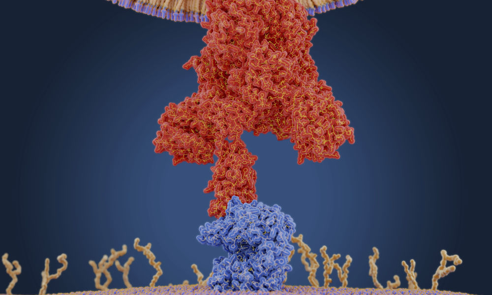
A coronavirus uses a protein on its membrane—shown here in red in a molecular model—to bind to a receptor—shown in blue—on a human cell to enter the cell. Once inside, the virus uses the cells' machinery to make more copies of itself. (Juan Gaertner / Science Source)
635
Rochester research into RNA structure and function provides key information for developing coronavirus treatments.

Viruses like the coronavirus that causes COVID-19 are able to unleash their fury because of a devious weapon: ribonucleic acid, also known as RNA.

A contingent of researchers at the University of Rochester study the RNA of viruses to better understand how RNAs work and how they are involved in diseases. As COVID-19 continues to spread around the globe, this research provides an important foundation for developing antiviral drugs, vaccines, and other therapeutics to disrupt the virus and stop infections.

Coronavirus update

The University’s website is a way to find guidance and critical information during a rapidly changing situation.

COVID-19 symptoms or exposure?

Find out what to do if you or a close contact have symptoms or think you may have been exposed.

“Understanding RNA structure and function helps us understand how to throw a therapeutic wrench into what the COVID-19 RNA does—make new virus that can infect more of our cells and also the cells of other human beings,” says Lynne Maquat, professor of biochemistry and biophysics at the University of Rochester Medical Center and the director of Rochester’s Center for RNA Biology.

In the past few decades, as scientists came to realize that genetic material is largely regulated by the RNA it encodes, that most of our DNA produces RNA, and that RNA is not only a target but also a tool for disease therapies, “the RNA research world has exploded,” Maquat says. “The University of Rochester understood this.”

In 2007, Maquat founded the Center for RNA Biology as a means of conducting interdisciplinary research in the function, structure, and processing of RNAs. The center involves researchers from both the River Campus and the Medical Center, combining expertise in biology, chemistry, engineering, neurology, and pharmacology.

While much of the research across the University has been put on pause, labs that are involved in coronavirus research remain active.

“Our strength as a university is our diversity of research expertise, combined with our highly collaborative nature,” says Dragony Fu, an assistant professor of biology on the River Campus and a member of the Center for RNA Biology. “We are surrounded by outstanding researchers who enhance our understanding of RNA biology, and a medical center that provides a translational aspect where the knowledge gained from RNA biology can be applied for therapeutics.”

How does coronavirus infect humans?

In mammals, such as humans, DNA contains genetic instructions that are transcribed—or copied—into RNA. While DNA remains in the cell’s nucleus, RNA carries the copies of genetic information to the rest of the cell by way of various combinations of amino acids, which it delivers to ribosomes. The ribosomes link the amino acids together to form proteins that then carry out functions within the human body.

Many diseases occur when these gene expressions go awry.

What does RNA stand for?

RNA stands for ribonucleic acid.

What is RNA?

RNA delivers the genetic instructions contained in DNA to the rest of the cell.

COVID-19, short for “coronavirus disease 2019,” is caused by the novel coronavirus SARS-CoV-2. Like many other viruses, SARS-CoV-2 is an RNA virus. This means that, unlike in humans and other mammals, the genetic material for SARS-CoV-2 is encoded in ribonucleic acid. The viral RNA is sneaky: its features cause the protein synthesis machinery of our cells to mistake it for RNA produced by our own DNA.

While SARS-CoV-2 is a new coronavirus, “it likely replicates and functions similar to related coronaviruses that infect animals and humans,” says Douglas Anderson, an assistant professor of medicine in the Aab Cardiovascular Research Institute and a member of the Center for RNA Biology, who studies how RNA mutations can give rise to human disease.

graphic created by the New York Times illustrates how the coronavirus that causes COVID-19 enters the body through the nose, mouth, or eyes and attaches to our cells. Once the virus is inside our cells, it releases its ribonucleic acid. Our hijacked cells serve as virus factories, reading the virus’s ribonucleic acid and making long viral proteins to compromise the immune system. The virus assembles new copies of itself and spreads to more parts of the body and—by way of saliva, sweat, and other bodily fluids—to other humans.

“Once the virus is in our cells, the entire process of infection and re-infection depends on the viral RNA,” Maquat says.

How is Rochester’s RNA research applicable to COVID-19? 

Horizontal portraits of Doug Anderson, Dragony Fu, and Lynne Maquat, scientists who study RNA of viruses.

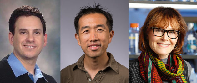
Researchers Douglas Anderson, Dragony Fu, and Lynne Maquat are among the scientists at the University of Rochester who study the RNA of viruses to better understand how RNAs work and how they are involved in diseases. (University of Rochester photos / Matt Wittmeyer / J. Adam Fenster)

Maquat has been studying RNA since 1972 and was part of the earliest wave of scientists to realize the important role RNA plays in human health and disease.

Our cells have a number of ways to combat viruses in what can be viewed as an “arms race” between host and virus. One of the weapons in our cells’ arsenal is an RNA surveillance mechanism Maquat discovered called nonsense-mediated mRNA decay (NMD).

“Nonsense-mediated mRNA decay protects us from many genetic mutations that could cause disease if NMD was not active to destroy the RNA harboring the mutation,” she says.

Maquat’s discovery has contributed to the development of drug therapies for genetic disorders such as cystic fibrosis, and may be useful in developing treatments for coronavirus.

“NMD also helps us combat viral infections, which is why many viruses either inhibit or evade NMD,” she adds. “The genome of the virus COVID-19 is a positive-sense, single-stranded RNA. It is well known that other positive-sense, single-stranded RNA viruses evade NMD by having RNA structures that prevent NMD from degrading viral RNAs.”

Maquat’s lab is currently collaborating with a lab at Harvard University to test how viral proteins can inhibit the NMD machinery.

Like Maquat, Fu studies fundamental aspects of RNA—and has found that his research on proteins may, too, be applicable to coronavirus research.

Fu’s lab analyzes enzymes and proteins that modify the chemical structure of RNA and how these chemical modifications impact the function of RNA. A research group at the University of California, San Francisco, recently identified an interaction between a protein made by the SARS-CoV-9 virus and a protein Fu studies.

“This is an intriguing result, and we are currently thinking of ways this interaction could affect the host cell,” Fu says. “There is emerging evidence that RNA-based viruses undergo RNA modification, so we could use this knowledge to identify key links between the host and pathogen for development of a coronavirus vaccine or treatment.”

How can RNA research be used to develop coronavirus treatments and vaccines?

One of the reasons viruses are such a challenge is that they change and mutate in response to drugs.

“Targeting viral RNA, or the proteins it produces, is key for treating this disease.”

That means novel virus treatments and vaccines have to be created each time a new strain of virus presents itself. Armed with innovative research on the fundamentals of RNA, scientists are better able to develop and test therapeutics that directly target the RNAs and processes critical to a virus’s life cycle.

The University of Rochester Medical Center, for instance, is currently participating in a clinical trial to evaluate the safety and efficacy of a potential coronavirus treatment called remdesivir, an antiviral drug particularly tailored to attack RNA viruses. The drug inhibits RNA polymerase, an enzyme responsible for copying a DNA sequence into an RNA sequence.

The Medical Center is additionally collaborating on a clinical trial to test the efficacy of an RNA vaccine. Traditional vaccines against viruses like influenza inject the body with inactivated virus proteins called antigens. The antigens stimulate the body’s immune system to recognize the specific virus and produce antibodies in response, with the hope that these antibodies will fight against future virus infection.

RNA vaccines, on the other hand, do not introduce an antigen, but instead inject a short sequence of the virus’s RNA. This messenger RNA provides cells with instructions to produce the virus antigens themselves. While no RNA vaccines have yet been approved in advanced vaccine trials, these vaccines have an advantage over traditional vaccines in that they would be easier and quicker to produce, and would allow the body to generate a more natural response to a virus.

Anderson has found that alternative therapeutics, such as the gene-editing technology CRISPR, may additionally “usher in a new approach to how we target and combat infectious diseases,” he says.

For the past few years, Anderson’s lab has developed tools and delivery systems that use the RNA-targeting CRISPR-Cas13 to treat human genetic diseases that affect muscle function. CRISPR-Cas13 is like a molecular pair of scissors that can target specific RNAs for degradation, using small, programmable guide RNAs.

When the health crisis first became apparent in Wuhan, China, researchers in Anderson’s lab turned their focus toward developing a CRISPR-Cas13 therapeutic aimed at SARS-CoV-2. Applying the knowledge already available about coronavirus RNA replication, they designed single CRISPR guide RNAs capable of targeting every viral RNA that is made within a SARS-CoV-2 infected cell. Using a novel cloning method developed in Anderson’s lab, multiple CRISPR guide-RNAs could be packaged into a single therapeutic vector (a genetically engineered carrier) to target numerous viral RNA sites simultaneously. The multi-pronged targeting strategy could be used as a therapy to safeguard against virus-induced cell toxicity and prevent ‘escape’ of viruses which may have undergone mutation.

“Infectious viruses and pandemics seemingly come out of nowhere, which has made it hard to rapidly develop and screen traditional small molecule therapeutics or vaccines,” Anderson says. “There is a clear need to develop alternative targeted therapeutics, such as CRISPR-Cas13, which have the ability to be rapidly reprogrammed to target new emerging pandemics.”

While many new treatments for the novel coronavirus are being considered, there is one thing that is certain, Maquat says: “Targeting viral RNA, or the proteins it produces, is key for treating this disease.”

Read more

person in protective gear examining something he is holdingNew URMC coronavirus research examines immune response
Researchers at the New York Influenza Center of Excellence are launching a new study to determine if and when a person could be re-infected with the novel coronavirus and whether some people have pre-existing immunity.
nanoparticle filter.Rochester researchers pursue quick ways to detect COVID-19
Nanomembranes, optical sensors, blood analysis—Rochester faculty are turning previous research avenues to focus on ways to quickly detect novel coronavirus to speed treatment.
Planet earth with a medical mask.Ethicists: COVID-19 pandemic a ‘wake-up call’
Rochester philosophy faculty explore moral dilemmas presented by the crisis and how they intersect with larger structural questions.


Tags: 

CategoryScience & Technology

Astra Zeneca has voluntarily paused this study. Why?


Update – September 9, 2020:  AstraZeneca has voluntarily paused this study pending the investigations of independent safety review committees in the U.K. and U.S.
2020-09-03_COVID_vaccine_432
Volunteer Daryl Moorehead, 63, of Rochester, NY, receives a nasal swab from project coordinator Sarah Northup. Photo by J. Adam Fenster

The University of Rochester Medical Center (URMC) is joining a phase 3 clinical trial for a potential coronavirus vaccine being developed by AstraZeneca and the University of Oxford known as AZD1222. 

This is the second phase 3 coronavirus vaccine study to be conducted in Rochester.  On July 27, four volunteers in Rochester were the first in the nation to receive an experimental vaccine being developed by Pfizer and BioNTech.  Rochester was one of only four sites in the nation that also participated in early stage studies of the Pfizer/BioNTech vaccine.  Phase 3 represents the final stage of human testing prior to regulatory approval, production, and mass distribution. 

The Rochester arm of the AstraZeneca study is being led by Ann Falsey, M.D., Angela Branche, M.D., Mike Keefer, M.D., and Catherine Bunce, B.S., M.S.  Falsey and Branche oversee the URMC Vaccine and Treatment Evaluation Unit and Keefer and Bunce lead the URMC HIV Vaccine Trials Unit.  Both programs are part of the national COVID-19 Prevention Network (CoVPN), which was formed by the National Institute of Allergy and Infectious Diseases (NIAID) to help lead the scientific response to the pandemic.  NIAID is headed by Anthony Fauci, M.D.  

The vaccine being developed by the British and Swedish company AstraZeneca and the University of Oxford uses a harmless adenovirus that contains the genetic material of the COVID-19 spike protein. The vaccine stimulates production of the surface spike protein, which primes the immune system to recognize the virus if infected. 

Phase 1/2 studies conducted in the U.K. – the results of which were reported on July 20 in the journal Lancet – found that the vaccine was not only safe, but generated an immune response to the virus.  Whether or not the vaccine provides protection from coronavirus infection across a wide range of age groups and medical conditions are questions that the phase 3 study will now seek to answer. In addition to the U.S., phase 3 studies of the vaccine are also under way in the U.K., Brazil, and South Africa. 

2020-09-03_COVID_vaccine_508
Volunteer Marie Kennedy of Rochester receives a dose of COVID-19 vaccine or placebo administered by Ian Shannon, RN. Photo by J. Adam Fenster

The AstraZeneca trial is funded by NIAID and the Biomedical Advanced Research and Development Authority (BARDA), part of the U.S. Department of Health and Human Services’ Office of the Assistant Secretary for Preparedness and Response. The trial is being implemented as part of the Operation Warp Speed, a multi-agency collaboration led by HHS that aims to accelerate the development, manufacturing and distribution of medical countermeasures for COVID-19.

The randomized placebo-controlled clinical trial will recruit 30,000 people across 81 sites in the U.S. including 1,000 volunteers in Rochester.  Researchers are focusing on individuals in the Rochester area ages 18 to 85 who are at greater risk for coronavirus infection, such as health care workers, first responders, teachers, and people who work in restaurants or retail.  Because COVID-19 has had a disproportionate impact of people of color, researchers are working with community partners to invite Black and Latinx individual to participate in vaccine trials. 

Individuals interested in volunteering for the study can visit www.covidresearch.urmc.edu or call (585) 276-5212.  

Wednesday, September 9, 2020

Dear Justin Trudeau: Prime Minister of Canada.

When a few dozen Mexican farm workers finally decide to avoid being exploited and decide to walk away from the farm fields of Canada, thousands of hungry college and university trained Canadian bureaucrats will wonder what went wrong with the economic system. They may also wonder how to grow vegetables in office buildings and yes, it is possible to grow vegetables in office buildings using hydroponic gardening methods, and that is what they should be doing instead of button pushing. 

 Millions of bureaucrats certainly don't know how or where to grow and harvest vegetables and the private multi-million dollar sky scraper office buildings they inhabit today, during daylight hours, lock their doors at night. Technically trained office workers are seldom valued. They work for large multi-national  exploitive companies who have CEO's willing to do anything to maintain the status quo...and that includes keeping office workers from climbing up the hierarchy ladder. Those same companies will lobby the Canadian government for tax breaks.

 While millions of office workers remain trapped in their offices, some farmers in Quebec, Canada, plow under their crops because not enough Mexican workers are available to help them harvest their vegetables. Other farmers dump their milk or kill their chickens if the market under values their produce.  What in hell is going on? Are Canadian students and welfare recipients not given the opportunity to work on the land? Why are there so many office bureaucrats and not enough Canadian born farm workers? Why are our educational priorities so screwed up? Why not send teenagers into the woods to plant trees and also to the farms to learn from the farmers and also, from the Mexican farm workers? I also suggest the Canadian government not subsidize Oil companies or any company that creates pollution and keeps thousands of office workers busy finding ways to manipulate the citizens of Canada.

---------------------------------------------------------

The response to the COVID-19 pandemic has shown what Canada can do when we treat a crisis like a crisis. We have thrown out the old rulebook of what is politically possible and focused on what is politically necessary to protect ourselves and our loved ones. 

I want to thank you for your work in these difficult times, but also to tell you that I don’t want us to go back to a ‘normal’, or a system which creates rampant inequalities based on a model of infinite exploitation and destruction of nature.

Now that the time has come to rebuild our future — and we will — we need to have courage and foresight to create something better. The recovery from COVID-19 must make our society more resilient, fair and sustainable. 

People in Canada deserve a world that values cooperation and caring and a government that embeds these values in our public policies. That respects nature and lives within its limits. A Canada where Indigenous rights and wisdom are not just a slogan, but protected by law. A world that recognizes that solutions to climate change can create great jobs and a better future.  

A green recovery for Canada means investing in: Trees and gardens and Green-Houses for every citizen in proportion to the territory they inhabit.

A much faster and just transition to a sustainable low-carbon economy if climate change does not destroy us first with viral pandemics and floods and forest fires!

The protection and restoration of land, freshwater, and ocean ecosystems along with the wildlife that call these places home is now next to impossible today, in 2020, with our present human population growth. Sex education in the schools must become a priority and the school buildings themselves must be transformed into large round Sunlit Green Houses with computers and quiet rooms to study Botany and Biology and Science.

The current land development companies creating Condos and Apartments and Industrial Parks everywhere there is an inch of space must be shut down and reimagined. They have become dangerous exploiters and are potentially creating future overcrowded slums! Developers are buying up every piece of farm land and small Green space they could find available in Canada and especially in Quebec, which considers itself an independent country. These money hungry developers are backed by our tax hungry municipal governments and the over-crowding they are creating, will cost the federal government more in the future, if there is a future, as global warming continues to heat up the planet and continues to change the atmosphere. A few years ago in countries such as Syria, over-crowding and poverty and misery created civil disobedience which lead to...war! I suggest we create housing apartments under farm lands using protected fibre optic cables to bring sunlight down to the people. We can also convert under-used industrial parks back into apartment buildings and wrap the buildings in Greenery. Literally create buildings with thousands of plants growing on the roof tops and inside and outside the buildings. Europe is ahead of us in this respect. 

An end to the use of single-use plastics, and the growth of a circular economy. Money is not edible!

The replacement of toxic chemicals used in agriculture and in the creation of consumer goods. We need cleaner and safer manufacturing alternatives. Elon Musk and his Gigafactory is an example of a clean manufacturing plant. 

The development of accessible and affordable and healthy communities and transportation networks.  For example the Electric commuter busses presently used in the city of Moscow, Russia.

A future that prioritizes well-being and social and racial justice with economic equity for all people living in Canada...created in partnership with Indigenous Peoples and the communities most exposed to environmental harm.

I urge you to speak out in favour of a green and just recovery with your fellow Parliamentarians and the media and I look forward to working with you and our community to build this better world. Thanks for reading

Sincerely,

SIGNED: JOSEPH NELSON RAGLIONE.

human4us2.com

Saturday, September 5, 2020

How and why do steroids workSteroids work by decreasing inflammation and reducing the activity of the immune system. Inflammation is a process in which the body's white blood cells and chemicals can protect against infection and foreign substances such as bacteria and viruses.Jan 20, 2020

 Gentle People:

  I am not a medical expert but as a journalist looking for important facts on how Covid-19 infects
and kills people, I often ask the question how can this virus be stopped? I may be wrong but I am
beginning to understand that there is a connection between an over-reactive immune system and the death of many seriously infected elderly people.
 I am learning that steroids work to decrease inflammation and they reduce the body's immune
 reaction to the SARSCoV2. Twenty percent of patients seriously  infected with Covid-19 
were saved from death by the medical use of steroids which basically boosted their hormones. 

 This is one more piece now in place to a large puzzle that has been plaguing us...excuse the pun!
Young people with more hormones are less likely to fall sick with SARSCov2 than their elders who
have lost hormones due to the aging process. It is now known that healthy and hormonal young
people are better protected from Covid-19 than their elders and the question is,  how do hormones 
and steroids work to protect both young and old from Covid-19? 

Attention research scientists, please explain why steroids are saving lives.

  Hello my good friend Valdemar Oliveira! I am happy to hear you had a successfull heart operation.  I hope you live to be 110. I may not be...